
Equine Health Library
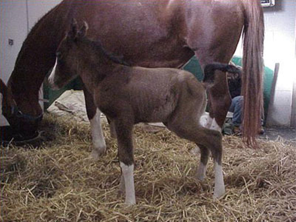
Foal
Health Conditions & Concerns
Neurologic | Gastrointestinal | Respiratory | Musculoskeletal
Neurologic
Neurologic Maladjustment Syndrome
Neonatal Maladjustment Syndrome is the most common condition that occurs as a result of oxygen deprivation. It is also referred to as dummy foal syndrome, peripartum asphyxia syndrome or hypoxic ischemic encephalopathy. Fortunately, the outcome for this condition is fair to good – the majority of these foals recover and grow to normal adults.
Neonatal maladjustment syndrome is caused by lack of oxygen and/or disruption of blood flow to the fetus before or during delivery. Some periparturient events that have been associated with the birth of dummy foals include severe systemic illness in the dam, premature placental separation (red-bag delivery), dystocia (difficult delivery due to abnormal positioning of the foal or excessive size of the foal), placentitis, induced delivery and cesarean section.
Many abnormal behavioral signs occur and are associated with varying degrees of brain edema and cell death.
Signs
- Jittery behavior
- Forgetting how and where to nurse
- Inability to follow the dam
- Bumping into walls as though blind
- Stuporous and sleepy behavior
- Seizures
- Coma
Other organ systems also may show damage due to lack of oxygen. Signs include abnormal intestinal motility, bloody diarrhea, decreased urine production and changes in lung function.
Treatment
- “Madigan Squeeze Technique” (MST) or “Madigan thoracic squeeze”
- Seizure control
- Treatments designed to minimize brain damage and preserve brain cell metabolism and nutrition
- Nutritional support and fluid therapy
- Immune system support, especially if the dummy foal is unable to ingest colostral antibodies
- Vigilant nursing care to be certain affected foals do not injure themselves.
Gastrointestinal
Gastrointestinal
Every foal should pass its first manure, or meconium, within 12 to 24 hours of delivery. Meconium is pasty or pelleted in consistency and dark brown or black in color. Most foals will strain a bit to pass meconium.
Meconium
Some foals, especially colts, may be unable to pass all the meconium, which results in a meconium impaction. Affected foals will flag their tails and strain repeatedly to pass manure. If this condition is not resolved early, it can lead to more violent colic accompanied by marked abdominal distention – the result of gas and fluid accumulating behind the impaction.
Colicky foals will stop nursing, and that begins a downhill spiral. A prophylactic enema administered shortly after birth helps reduce the risk of meconium impaction. Following meconium passage, the foal’s feces should be soft and light tan in color.
Foals should pass meconium, or first manure, within 12 to 24 hours of delivery
Colostrum
Colostrum ingestion is critical for many reasons, including passive transfer of antibodies to protect your foal from viral and bacterial diseases. Mares produce colostrum during the last two to five weeks of pregnancy, and good colostrum is a result of good nutrition and a sound vaccination program.
Colostrum is a rich source of calories, vitamins A and E, white blood cells, growth factors and other hormones, and enzymes. Laxative properties also have been ascribed to its ingestion. The predominant antibody in equine colostrum is IgG.
The quality of colostrum varies between mares, and older mares may have poorer quality colostrum. If your mare begins to leak colostrum hours or days prior to delivery, she is losing the richest source of antibodies. If you observe your mare leaking colostrum, contact your veterinarian. Remedies include saving that colostrum that is being lost and bottle feeding it back to your foal immediately after birth and/or using a commercially available test kit to measure the quality of your mare’s colostrum after delivery. If necessary, your veterinarian can provide you with an alternate source of antibodies for your newborn foal.
Colostral antibody concentrations decline rapidly to negligible levels within 12 hours of delivery in mares that are being nursed regularly by healthy foals. These antibodies provide “passive” immunity for your foal for the next three to five months.
Your foal should ingest at least 0.5-1.0 L (16 to 32 oz.) of good quality colostrum within the first 8 to 12 hours life to ensure absorption of adequate antibodies. Peak absorption occurs during the first three to six hours following birth. The primary antibody in colostrum is IgG.
Healthy foals that have nursed and absorbed adequate colostrum should have an IgG concentration in their bloodstream of at least 800 mg/dl. Your veterinarian can draw a blood sample from your foal within 12-24 hours of delivery and can quickly and accurately measure the IgG concentration.
Newborn foals with IgG concentrations less than 400 mg/dl are said to have “failure of passive transfer” and should receive supplemental colostrum and/or a plasma transfusion to provide vital antibodies that help reduce the risk of serious bacterial and viral infections during the first few months of life. A complete blood count also can be performed on each newborn foal to detect early signs of infection or anemia.
You should observe your newborn foal frequently during the first few weeks of life to detect early signs of disease. Often the first sign of a sick foal is lethargy, sleeping more than usual and decreased nursing vigor accompanied by an overly distended udder on the mare. Young foals are at risk for a variety of respiratory diseases and diarrhea. Monitor your young foal’s breathing rate and effort, body temperature, nursing behavior and manure consistency.
Common causes of newborn foal illness
- Overwhelming bacterial infection (septicemia)
- Prematurity or dysmaturity
- Neonatal maladjustment syndrome (neurological dysfunction associated with lack of oxygen before or during delivery)
Diarrhea
There are multiple causes of diarrhea that may affect your foal. Treatment can vary depending on the underlying cause, the severity of the diarrhea and the age of the foal.
Foal heat diarrhea
Foal heat diarrhea occurs in foals between one and two weeks of age, typically when the mare is experiencing her first heat or estrous cycle, called foal heat. Originally it was thought that foal heat diarrhea was related to hormonal changes in the mare’s milk composition, however even orphaned foals develop diarrhea. Now, veterinarians believe it is a transitory, age-related diarrhea caused by changing bacterial flora within the foal’s intestines.
Foals with foal heat diarrhea are not systemically ill. Affected foals do not have a fever and usually remain bright and continue to nurse. The diarrhea is usually mild, slightly loose in consistency and self-limiting. Affected foals rarely require medical treatment. Younger foals and those with more severe diarrhea can be an exception and may require treatment with fluid therapy, anti-diarrheal medication and probiotics.
Rotavirus A and B diarrhea
Rotavirus is a common, highly contagious cause of diarrhea in young foals on large breeding operations. Foals are infected through contact with manure from other infected foals.
The incubation period (time from contact with the virus to onset of clinical signs) is a short 12 to 24 hours. The average age of foals affected by rotavirus is two to three months, but foals from two days to five months can become infected. The younger the foal, the more severe the signs. Large numbers of susceptible foals on a farm can become infected, but fortunately mortality rates are low. The virus infects absorptive cells in the foal’s small intestine, which results in an inability to absorb milk and other nutrients.
Clinical signs vary in severity depending on the foal’s age and immune status
- Depression
- Lethargy
- Loss of appetite
- Diarrhea that can vary in consistency from “cow pie” to watery
Therapy is symptomatic and includes intravenous fluids to correct electrolyte imbalances and administration of anti-diarrheal and gastroprotectant medications. Pain control may be required for those foals exhibiting mild colic. Anti-ulcer medication is often administered.
A Rotavirus A vaccine is commercially available for use on farms with a history of rotavirus infections. This vaccine is administered to pregnant mares at eight, nine and 10 months of gestation to improve colostral immunity. This series of three pre-foaling vaccinations must be repeated for every pregnancy. Rotavirus B vaccine is not available.
Salmonella diarrhea
Salmonella diarrhea is one of the most serious causes of diarrhea in young foals. Salmonella is a gram-negative bacterium. Foals that have been given antibiotics, have a suppressed immune system or have had abdominal surgery are more predisposed to infection.
In a healthy foal, normal gastrointestinal flora inhibit the proliferation and colonization of Salmonella sp. by competing for available space and nutrients and by secreting bacteriocins (e.g., short chain fatty acids) that are toxic to Salmonella. Normal gut motility helps prevent overgrowth of bad bacteria. The most likely source of infection for foals is another horse, perhaps an asymptomatic shedder, or even their dams.
Signs of Salmonella diarrhea
- Mild to severe and possibly bloody diarrhea
- Rapid dehydration accompanied by hypoproteinemia (low protein concentrations in the blood)
- Fever
- Loss of suckle
- Colic
- Toxic mucous membranes
- Septicemia
Salmonella diarrhea in young foals is often associated with sepsis and can be accompanied by extra-intestinal infections including osteomyelitis (bone infection), arthritis, pneumonia, peritonitis, cellulitis and nephritis (kidney infection).
Blood work often reveals a low white blood cell count, low protein concentration and marked electrolyte changes. Diagnosis is based on serial fecal cultures and/or PCR testing of feces obtained daily for three to five days.
Treatment involves aggressive IV fluid therapy, hyperimmune plasma administration, pain control, gastroprotectants, anti-diarrheal medications and broad-spectrum bactericidal antibiotics. Good nutrition is essential. Strict hygiene is paramount to control spread of the infection.
Phenolic compounds (O-Syl, One Stroke Environ) and dilute bleach are effective disinfectants following removal of all organic debris from surfaces to be disinfected. More recently, Virkon S®, a peroxygen disinfectant, has been shown to be more effective than the quaternary ammonium disinfectants when used in footbaths to reduce bacterial contamination on footwear. Virkon is biodegradable and has low toxicity.
Affected foals should not be reintroduced to the general population until five consecutive fecal samples are negative for Salmonella.
Salmonella is not only a threat for your foal but is also a potential human pathogen. Work closely with your veterinarian not only to provide proper care for your foal but also to institute aggressive biosecurity measures to protect other foals and horses on the premises and all human personnel working with infected individuals.
Clostridial diarrhea
Clostridial enteritis has been increasing in frequency. C. perfringens (most commonly Type A, less commonly Type C) and C. difficile (toxigenic strains produce toxins A and B) have both been identified as causative agents of colic and diarrhea in young foals as young as one day of age.
Clostridial enteritis can be sporadic or can affect groups of foals on a farm when Clostridial spores are ingested from the environment. Foals may be more susceptible to infection and overgrowth of the bacteria within the gut due to several factors including: Immaturity of the bile, failure of passive transfer, ingestion of diets high in carbohydrates and protein, hypoxic gut damage, concurrent infection with other pathogens such as rotavirus and concurrent administration of antibiotics.
During Clostridial enteritis, the small intestine is usually more severely affected than the large intestine. Diffuse mucosal edema with localized damage to the tips of villi or sloughing of the entire villous epithelium has been observed. Loss of the protective mucosal lining contributes to the development of endotoxemia and passage of bacteria from the gut lumen into the foal’s bloodstream.
Clinical signs of Clostridial diarrhea
- Colic, which can be severe
- Mild to moderate abdominal distention
- Diarrhea (Diarrhea associated with C. perfringens is often bloody; diarrhea due to C. difficile may be bloody but can also be brown and fetid)
- Fever is usually present
Diagnosis is based on farm history, history of concurrent use of antibiotics, presence of fever and low white blood cell count. Therapy includes the symptomatic treatments described above and pain control. The antibiotic of choice is metronidazole. Severe cases also should receive broad-spectrum antimicrobial therapy. One to two liters of hyperimmune plasma are beneficial.
Additional therapy includes aggressive IV fluid support. Anti-ulcer medications are usually indicated. Probiotics also may be helpful. Foals that are very colicky with marked abdominal distention should be prevented from nursing for a while and supported with intravenous (parenteral) nutrition. Some foals recovering from Clostridial enteritis have displayed varying degrees of lactose intolerance and benefit from administration of commercially available Lactaid tablets.
Lawsonia: Equine Proliferative Enteropathy (EPE)
Caused by Lawsonia intracellularis, EPE is an emerging enteric disease most commonly documented in foals and weanlings between two and eight months of age. Possible risk factors include the decline of maternal antibodies and the stress of weaning.
Clinical signs of Lawsonia
- Diarrhea accompanied often by profound weight loss
- Anorexia (loss of appetite)
- Lethargy
- Depression
- Ventral and limb edema due to hypoproteinemia (low concentration of protein in the blood stream)
Lawsonia infection causes a secretory diarrhea and is associated with small intestinal mucosal thickening and proliferation. The diarrhea is typically acute but may be intermittent in some cases and can vary in consistency from soft feces to profuse watery diarrhea. The most consistent clinicopathological abnormality is hypoalbuminemia (a small protein in the blood).
Transabdominal ultrasonography typically reveals an increase in small intestinal wall thickness. Successful therapy includes intravenous plasma administration to correct the hypoproteinemia and antibiotics to treat the underlying infection.
Gastroduodenal Ulcer Disease
Ulcers can develop in the stomach and small intestine of growing foals.
Signs of Ulcers
- Diarrhea
- Loss of appetite
- Teeth grinding
- Rolling up on the back in a cast position
- Low-grade colic
- Treading
- Salivation
- Many foals become colicky shortly after nursing
- Occasionally manure from affected foals will be occult blood positive
Ulceration of the duodenum (a segment of the small intestine) can result in narrowing of the intestine as ulcers begin to heal, leading to stricture formation and obstruction. Foals with this condition develop a backup of ingesta into their stomachs, which can result in colic and spontaneous reflux of stomach contents out their nose. Ulcers in the duodenum can also predispose to an ascending infection that travels up the bile ducts to the foal’s liver, resulting in hepatitis.
Diagnosis and treatment
Your veterinarian will diagnose ulcers based on clinical signs and examination of the foal’s stomach lining with a gastroscope. Duodenal stricture formation is best visualized with contrast radiography.
Foals at highest risk for ulcers are foals suffering from hypoxic gut damage, infectious diarrhea, painful orthopedic conditions and excessive use of certain non-steroidal anti-inflammatory drugs such as phenylbutazone. The stress of weaning also may increase the risk of ulcer formation.
Therapy includes the use of various anti-ulcer medications. Your veterinarian will prescribe a suitable treatment plan for your foal.
Respiratory
Respiratory
In the very young foal, pneumonia is frequently part of a generalized septicemic condition. In the older foal, it often presents as a single organ infection. Bacterial pathogens invade the lungs via inhalation or via the blood stream. Pneumonia may be acquired in utero, during the birth process or, more frequently, during the weeks and months following delivery.
Bacterial pneumonia
Bacteria responsible for foal pneumonia are often the same pathogens responsible for neonatal septicemia and include gram-negative enteric pathogens: E. coli, Klebsiella, Actinobacillus, Pasteurella and Salmonella. Beta-hemolytic Streptococcus sp (S. zooepidemicus) are the most common gram-positive organisms involved.
Foals suffering from difficulty swallowing (dysphagia) are prone to develop aspiration pneumonia. Causes of dysphagia include upper airway anomalies, White Muscle Disease, botulism and dysmaturity/prematurity. Ascarid larval migration through the lungs also can contribute to persistent, low-grade lung inflammation and predispose the foal to recurrent episodes of infectious respiratory disease. Older foals may develop a very specific bacterial pneumonia caused by Rhodococcus equi.
Viruses capable of causing respiratory disease in young foals include Equine Herpesviruses and, less commonly, Equine Adenovirus. Equine Influenza virus typically does not affect young foals and weanlings but can be a problem among yearlings and 2-, 3-, and 4-year-old youngsters.
Reactive airway disease or allergic lung disease also occurs in the older foal and can be confused with pneumonia.
Foals with pneumonia can have a wide spectrum of clinical signs
- Mild nasal discharge
- Intermittent cough
- Profuse, purulent nasal discharge
- Persistent cough
- Respiratory distress
- Fever
- Loss of appetite
- Resting respiratory rates greater than 40 breaths per minute in older foals
Nasal discharge and cough are relatively uncommon signs in the newborn foal and become more reliable indicators of respiratory disease as the foal ages. Increased breathing rate and exaggerated respiratory effort are also good indicators of underlying lung disease. Exercise often exacerbates signs of respiratory disease, so watch your foals as they run and play.
Listening to the foal’s chest with a stethoscope can reveal abnormal lung sounds suggestive of lung disease.
Diagnosis and treatment
Your veterinarian may use a variety of tools to diagnose pneumonia. A complete blood count often reveals an elevated white blood cell count. Radiographs (X-rays) are particularly helpful in detecting lung lesions (areas of consolidation, abscesses) and their distribution. Thoracic ultrasonography is helpful in detecting early signs of lung consolidation and abscessation that occur along the outer surface of the lungs. Cultures of blood, tracheal aspirates, bronchoalveolar lavage (BAL) fluid or nasopharyngeal swabs help identify bacterial pathogens. PCR testing of nasal swabs can be used to identify viral infections. Broad-spectrum bactericidal antibiotic therapy is the cornerstone of treatment for pneumonia caused by bacteria.
Rhodococcus equi pneumonia
Rhodococcus equi (R. equi) causes a severe, often-fatal bronchopneumonia in foals less than six months of age.
R. equi pneumonia can be financially devastating for the owner as it involves expensive, long-term antibiotic therapy with drugs that have the potential for side effects (e.g., diarrhea and high, drug-related fevers) and a decreased value of “survivors” due to poor growth and unthriftiness.
Morbidity rates due to R. equi vary worldwide between 5 and 20 percent with mortality rates as high as 80 percent. Farms where R. equi is endemic experience much higher morbidity rates. Recovery rates also vary. Some studies show survival rates above 75 percent with more than 50 percent of those survivors eventually racing. Early detection and aggressive therapy is instrumental in improving survival.
R. equi is a gram positive, rod-shaped bacterium that lives in the soil and is resistant to desiccation and sunlight. Because horse manure provides the simple organic acid substrates on which the organism thrives, there is progressive development of infection in the soil on well-established horse farms. Warm, dry conditions help the organism multiply and disperse. Unfortunately, there is no way to eradicate R. equi from the environment.
These bacteria can actively multiply within the intestines of foals less than three months of age. When foals with R. equi pneumonia cough up and swallow infected sputum, the disease-causing R. equi can multiply within the intestines. Feces from these infected foals can contain very high numbers of infective R. equi bacteria. Therefore, foals with R. equi pneumonia are a major source of pasture contamination. Under favorable conditions, a gram of contaminated soil on endemic farms can contain millions of virulent R. equi. Inhalation of dust laden with R. equi is the primary route of pneumonic infection.
Foals receive passive immunity against R. equi through colostrum, but maternally derived immunity diminishes by two months of age. Foals are not able to produce adult levels of protective antibodies until at least 5 to 6 months of age, which helps explain why foals are most susceptible to R. equi infection between one and five months of age. Genetic predisposition and other host-related factors may also play a role. Dry, dusty weather also may favor transmission.
Clinical signs of respiratory disease due to R. equi
- Elevated breathing rate (greater than 60 breaths per minute)
- Labored breathing
- High fevers (greater than 104 degrees Fahrenheit)
- Exercise intolerance
- Coughing is variable
- Other non-specific signs include lethargy, weight loss and unthriftiness
Some foals develop other clinical signs in addition to pneumonia. These signs include puffy joints, uveitis (inflammation within the eye), diarrhea and weight loss.
Diagnosis
Lab work may reveal a markedly elevated white blood cell count and fibrinogen concentration. Therapy includes oral antibiotic therapy using rifampin and one of the following drugs: Erythromycin, clarithromycin or azithromycin.
Treatment and Prevention
No vaccine is available, so a breeder’s attention should be focused on decreasing the risk of infection among newborn foals, recognizing early disease among infected foals and using new antibiotics to treat established disease.
Reducing the risk of infection begins with environmental management. Environmental control strategies for enzootic farms include decreasing dust formation on pastures and paddocks, housing foals in well-ventilated areas, rotating pastures, reducing the size of mare-foal bands, irrigating and planting dirt areas with grass, and removing feces frequently from stalls, paddocks, indoor arenas and pastures. Breeding mares earlier in the season to ensure foaling during colder weather may help reduce the number of susceptible foals exposed to dry, dusty summer conditions.
The best prevention on farms with an increased incidence of R. equi pneumonia is early intravenous administration of hyperimmune (HI) R. equi plasma to susceptible foals. Administration of HI plasma containing antibodies against various R. equi antigens has been effective in reducing the incidence and severity of R. equi pneumonia in foals.
Vaccinations against other respiratory pathogens should be kept current, and foals should be maintained on an aggressive deworming program to reduce heavy burdens of ascarid larvae in the lungs that can increase the risk of secondary bacterial infections, including R. equi.
Musculoskeletal
Musculoskeletal
Angular limb deformities can be caused by abnormal positioning within the uterus, genetics or congenital defects associated with an in-utero insult during pregnancy. Severe deformities may prevent the foal from standing and nursing.
Angular limb deformities
Any condition that keeps your foal down on the ground increases the risk of other conditions, including pressure sores, patent urachus, pneumonia and conditions associated with lack of adequate nutrition and critical antibodies. The pain associated with limb contractures can also increase your foal’s risk for gastric (in the stomach) or intestinal ulcers.
Your veterinarian will determine a treatment plan based on how severe the limb deformity is and which joints and limbs are affected.
Therapeutic options
- Physical therapy
- Casts, PVC splints, DynaSplint™, corrective shoes
- Oxytetracycline administration
- Conservative use of analgesics or painkillers (e.g., Banamine® (flunixin meglumine), phenylbutazone)
- Anti-ulcer therapy
- Controlled, conservative exercise to be certain that the affected foal does not become fatigued. Foals that over-exercise can make the deformities worse
If your foal has limb deformities, schedule regular veterinarian exams to make sure the deformity is improving in an appropriate manner and timely fashion. Your veterinarian may work closely with your farrier to design special shoes to help correct certain deformities.
Important Safety Information
Banamine: For Oral Use in Horses Only. Not for use in horses intended for human consumption. Do not use in horses showing hypersensitivity to flunixin meglumine. The effect of BANAMINE Paste on pregnancy has not been determined. Concomitant use of Banamine with other anti-inflammatory drugs such as NSAIDs and corticosteroids should be avoided or closely monitored. For complete information on Banamine® Paste, see accompanying product package insert.
Developmental Orthopedic Disease (DOD)
The term DOD typically includes the following conditions
- Osteochondritis dissecans: Development of a dissecting cartilage flap on the articular surface of the joint, which results in joint inflammation and pain
- Subchondral cystic lesions: Defects in the bone underlying the joint surface; most common locations are the stifle and fetlock joints
- Angular limb deformities: Deviation of the limb from a straight line; often involves bones below major joints such as the carpus, hock or fetlock; foals can be born with angular limb deformities or can develop deformities as they grow
- Physitis: Painful swelling around the growth plates at the end of the forearm (radius) and/or metacarpus (cannon bone)
- Flexural deformities: Angulation occurring around a joint; usually some form of contracture; fetlock, pastern and carpal joints are the most frequently involved; foals can be born with contracture deformities (i.e., congenital) or can develop these deformities during the first 18 months of life
- Abnormalities involving the cuboidal bones (small bones in the carpus and hock): Typically seen in premature or immature foals born with incompletely calcified cuboidal bones; in some foals, these bones calcify abnormally resulting in misshapen (usually wedge-shaped) bones that predispose the foal to arthritis in the affected joint
DOD can result from many different factors. Congenital forms of DOD can be detected during your newborn foal’s first veterinary examination within 24 hours of delivery. But many forms of DOD develop during the first year of life and initially may be subtle in appearance.
Regular veterinary exams during this time period help detect limb abnormalities early and begin corrective treatment that can prevent career-ending lamenesses. Conformation, genetics, diet and exercise regimens all play a role in DOD. Whenever your veterinarian visits for vaccinations or other routine procedures always include a lameness and conformation exam for your growing foals.