Equine Health Library
Pleasure Horse
Health Conditions
Neurologic | Gastrointestinal | Respiratory
Neurologic
Neurologic
If your horse’s balance seems off, or he is starting to stumble more often or seems uncoordinated, has a head tilt, or begins to cross canter rather than pick up a lead like he usually does, call your veterinarian right away.
A complete neurological exam can help identify the cause and plan additional diagnostics and therapy. For most diseases affecting the central nervous system (CNS), the earlier treatment can begin, the better the chances are for a full recovery.
Eastern Equine Encephalomyelitis (EEE, Sleeping Sickness)
Mosquitoes aren’t just annoying; they can also carry viruses that cause neurological diseases like Eastern Equine Encephalitis (EEE).
EEE is a virus that causes severe and often fatal neurologic disease in horses, humans, and other animals. The disease is found as far north as eastern Canada and as far south as Central and South America. In the United States, EEE is most commonly detected in the Atlantic and Gulf Coast states and the Great Lakes region. Every year there are reports of unvaccinated horses diagnosed with EEE.
Clinical Signs of EEE
- High fever
- Severe depression
- Incoordination
- Paralysis
- Seizures
- Coma
Unfortunately, EEE is nearly always fatal in horses. Prevention focuses on mosquito control and appropriate vaccination once or twice per year depending on the climate and other disease risk factors. The American Association of Equine Practitioners (AAEP) recommends that all equids (horses, donkeys, mules) get vaccinated against EEE at least annually. Since the virus is spread by mosquitoes, horses who live in areas with long mosquito seasons are typically vaccinated against EEE twice a year.
Rabies
The word rabies strikes fear into the horse owner’s heart for good reason. The disease is invariably fatal, has no treatment or cure, and poses a significant risk for human exposure. Thankfully, there are only a few human cases of rabies reported in the United States each year, but because there is no treatment or cure this disease must be prevented.
The most common wildlife reservoirs for rabies are skunks, raccoons, foxes, and bats – animals that are often seen around barns and pastures. The incubation period (time from bite to onset of signs) for rabies can range from days to months. The average survival time following onset of neurologic signs is five days. Multiple cases of equine rabies are reported every year and most of these horses are either known to have been unvaccinated or their vaccine history is unknown. For this reason, it is very important to vaccinate your horses every year against rabies.
Rabies can look like anything and everything. The clinical signs of rabies can be highly variable and vague, especially early in the course of disease.
Possible Clinical Sign of Rabies
- Lameness
- Colic
- Problems with genital and urinary tracts
- Central nervous system-specific signs including cranial nerve deficits (head tilt, difficulty swallowing)
- Bizarre behavior ranging from stupor to aggression and self-mutilation
Because there is no cure, rabies prevention focuses on annual vaccination. The American Association of Equine Practitioners (AAEP) recommends every horse, donkey, and mule be vaccinated against rabies once a year.
For more information about rabies, click here. Download the Rabies Quick Facts
West Nile virus (WNV)
A flavivirus transmitted by mosquitoes that can infect horses and humans, WNV was first diagnosed in U.S. horses in 1999 in the state of New York. Since that time, WNV has been detected in horses in all 48 continental United States, as well as most of Canada and Mexico. The severity and duration of clinical signs can vary greatly with WNV.
Signs of WNV
- Depression
- Low-grade fever
- Change in behavior
- Muscle fasciculations (twitching)
- Incoordination, ataxia
- Cranial nerve deficits (such as head tilt, ear droop, difficulty swallowing)
- Recumbency
Once a horse is infected and showing clinical signs, mortality rates can reach over 30 percent. And of the horses who do survive, not all of them experience a full recovery.
There is no treatment for the virus itself, just supportive care to help the horse recover on its own. Annual vaccination and mosquito control are the best methods of prevention. The AAEP recommends that all horses, donkeys, and mules be vaccinated against WNV at least once a year. Since the virus is spread by mosquitoes, horses who live in areas with long mosquito seasons are typically vaccinated against WNV twice a year.
Equine Protozoal Myeloencephalitis (EPM)
EPM is known to be caused by 2 different protozoans – Sarcocystis neurona and Neospora hughesi. The vast majority of EPM cases are caused by S. neurona. Opossums are the definitive host of S. neurona and they shed infective sporocysts in their feces. Horses are exposed to S. neurona by ingesting feed or water contaminated with feces from an infected opossum. In the eastern 2/3 of the US more than 80 percent of horses have been exposed to S. neurona and in the western US 68 percent have been exposed. Thankfully, only a small percentage of exposed horses develop neurologic disease. We don’t yet know why some horses get sick while most horses do not.
Risk factors include:
- Age, with mature horses more commonly affected than very young horses
- Career or lifestyle, with a higher incidence among horses used for racing or western performance
- Recent stress event such as long-distance transportation or illness
- Season, with increased numbers of cases identified during late summer and fall
- Management practices that include not protecting horse feeds from opossums and/or leaving pet food in the barn as an opossum attractant
- Barns and pastures in close proximity to wooded terrain
- History of other cases of EPM on the farm
Clinical Signs
Widely variable because they are dependent on the random path the protozoans take within the central nervous system (brain and spinal cord) where they cause inflammation and damage to the tissues
- Ataxia (wobbly, unsteady gait)
- Muscle atrophy – can be mild or severe
- Asymmetrical clinical signs
- Difficulty swallowing
- Head tilt
- Seizures
- Inability to stand
Diagnosis
Unfortunately, definitive diagnosis can only be confirmed by an autopsy. In a live horse, the most accurate diagnosis is achieved by comparing levels of antibodies against S. neurona and N. hughesi found in the blood to those antibody levels found in the cerebrospinal fluid (the fluid that surrounds the brain and spinal cord) in a “serum:CSF ratio” test.
Treatment
Fortunately, there are multiple FDA-approved medications available to treat EPM effectively. PROTAZIL® (1.56% diclazuril) Antiprotozoal Pellets is a safe and effective EPM treatment that comes in the form of alfalfa-based pellets. Your veterinarian will guide you on appropriate treatment for EPM.
Important Safety Information
PROTAZIL® is contraindicated in horses with known hypersensitivity to diclazuril. The safety of PROTAZIL® in horses used for breeding purposes, during pregnancy, or in lactating mares, and use with concomitant therapies in horses has not been evaluated. Do not use in horses intended for human consumption. Not for human use. For complete safety information, refer to the product label.
Gastrointestinal
Colic: The Dreaded 5-Letter Word
One of the most common gastrointestinal problems is colic. Because the term “colic” describes a clinical sign and is not a disease itself, the term has become a catchall for any type of abdominal pain. Some causes of colic can be fatal and colic is one of the leading causes of death in the horse. Fortunately, most colic cases are mild and respond well to medical therapy.
Causes
Abdominal pain associated with colic can be caused by a problem in any part of the gastrointestinal tract or by a problem in another internal organ, such as colic from liver disease, heart dysfunction, bladder or kidney disease, or disorders involving the reproductive organs.
Common Gastrointestinal Causes of Colic
- Ileus (lack of gut motility)
- Obstruction (due to foreign body)
- Impaction (obstruction due to feed/fecal material or parasites)
- Displacements or torsions (twists) of the small or large intestines
- Gas/spasmodic colic
- Parasites (especially roundworms in foals and tapeworms in adult horses)
- Infarctions (obstruction of blood supply)
- Ulcers in the stomach or intestines
- Idiopathic colic (colic of unknown cause)
Clinical Signs of Colic
Signs of colic depend on the severity of pain and include one or more of the following:
- Lying down more than normal or getting up and down repeatedly
- Standing in a stretched-out posture, urinating small volumes frequently
- Looking at or biting at the flank
- Sweating accompanied by restless, anxious behavior
- Circling, pawing, kicking out, rolling
- Abdominal distention
- Decreased manure production or diarrhea
- Elevated heart rate
- Elevated respiratory rate
- Loss of appetite
Here’s some good news: The vast majority of all colic cases respond to symptomatic medical therapy; only a small percentage require surgical intervention.
Causes of colic that are usually treated with medical therapy include: most impactions, gas/spasmodic colic, diarrhea, or enteritis (inflammation of the lining of the small intestines), most stomach ulcers and ileus. Gastrointestinal problems that require surgery include most cases of intestinal displacement, torsion of intestinal segments, severe impactions that are not responding to medical treatment with fluids and laxatives, and intestinal strangulations/obstructions.
Types of Colic
- Regional and diet-related colic:
- Enteroliths are hard, usually round concretions that can reach softball size or larger and become lodged in the large or small colon, creating an obstruction. This type of colic is more common among horses in certain regions of the country, such as California, where horses ingest alkalinizing feed high in protein, phosphorus, and magnesium (such as alfalfa) along with water that’s high in magnesium. Certain breeds – such as Arabians, Arabian crosses, Morgans and Miniature Horses – may be more predisposed to develop enteroliths. Surgery is required to treat this condition.
- Horses grazing and eating off sandy soil are at increased risk for sand impactions. Sand impactions can often be treated medically unless severe.
- Activity, travel/stress, and weather-related colic: Horses experiencing sudden decreases in activity, such as strict stall confinement following an injury or illness, are at greater risk for developing a feed impaction in the cecum or large intestines. The same concerns apply for horses who are traveling and/or stressed, although for slightly different reasons. Horses who travel and/or are stressed often do not consume as much water while traveling as they usually do at home, which results in dehydration and an increased risk of impaction colic. Decreased water consumption, dehydration, and subsequent impaction colic may also occur during cold winter months and hot summer months if adequate amounts of clean fresh water are not available. Most impactions can be treated with fluid therapy (administered orally and/or intravenously) and a variety of laxatives given via stomach tube. Occasionally, surgical intervention is required for refractory impactions.
- Parasite-related colic:
- Tapeworm infestations can cause spasmodic colic, impactions of the small intestine and intussusceptions involving the distal small intestine and cecum.
- Heavy ascarid (roundworm) burdens can cause small intestine impaction and even bowel rupture in foals and weanlings.
- Encysted small strongyles (cyathostomes) are the larval form of an intestinal parasite that encysts in the lining of the wall of the large intestine. These encysted larvae can cause significant inflammation of the gut wall when large numbers of hibernating larvae begin to emerge simultaneously, resulting in colic accompanied by weight loss and diarrhea.
- Medication-related colic: Horses receiving high-doses or chronic therapy with non-steroidal medications such as phenylbutazone are at increased risk for gastric and intestinal ulcers. Horses with gastric ulcers frequently present with colic and loss of appetite, while horses with ulceration of their large colon often present with colic and diarrhea.
Diagnosis and Treatment
Call your veterinarian immediately if you have concerns that your horse is showing signs of colic. Your veterinarian may want you to provide vital signs (heart rate/pulse, respiration, and rectal temperature) when you call. These parameters will help your veterinarian assess the severity of the colic and give you recommendations on what to do until he or she arrives to evaluate your horse.
What to do until your veterinarian arrives
- Check your horse’s heart rate. The normal range is 32 to 44 beats per minute
- Check your horse’s respiration rate. Normal resting breathing rate is 8 to 12 breaths per minute
- Take your horse’s temperature if it is safe to do so. Normal temperature range is 99 to 101.4 degrees Fahrenheit. The presence of a fever may indicate infection or inflammation somewhere in the body.
- Observe the amount and consistency of manure in the stall
- Observe any abnormalities in the amount of grain, hay or water consumed
- Be prepared to report all medications administered and any recent changes in diet or activity
- It is ok for your horse to lay down quietly
- If your horse is trying to roll while lying down, get him up and walking
- Remove all hay and grain, and prevent your horse from eating until the cause of colic is determined and your veterinarian has a plan of action
- Have a game plan in place in case your horse needs to be transported to a hospital for further treatment. It is also a good idea to let others know your recommendations and preferred treatment should your horse suffer from a serious colic episode, and you are unable to be reached
- If your horse is insured, understand the specifics of your policy and who to contact if necessary.

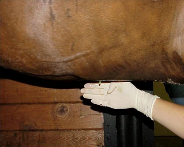
Peritoneal tap to obtain a sample of the fluid within the abdominal cavity and surrounding the intestines. Evaluation of the peritoneal fluid can help identify the cause of colic.
Diagnosing Colic
- Upon arrival, your veterinarian’s assessment may vary depending on the severity of signs
- Your veterinarian may begin with a history of your horse’s recent activity, diet and any medications administered
- Next, your veterinarian will likely perform an examination including evaluation of your horse’s heart rate, respiratory rate, body temperature, color of mucous membranes, hydration status and gastrointestinal sounds
- Rectal palpation is used to try and identify abnormalities of the intestinal tract such as impactions, gas or fluid-distended loops of intestines, enteroliths, intestinal twists or displacements
- If indicated, your veterinarian may pass a stomach tube down your horse’s esophagus and into the stomach to detect the presence of reflux (stomach and intestinal fluid that accumulates as a result of obstruction or loss of intestinal motility)
- A peritoneal tap may also be performed. This involves sterile placement of a cannula or needle into the abdominal cavity to obtain a sample of peritoneal fluid that bathes all the internal abdominal organs. There will be changes in the composition of peritoneal fluid if there are severely diseased segments of bowel.
- Ultrasound can also be used to evaluate the abdomen and gastrointestinal tract.
While most colic cases are able to be successfully treated medically, some horses with colic signs require surgical intervention for a successful outcome. It’s important to think about your preferences, transportation options, and financial resources prior to your horse experiencing an episode of colic. Thinking about these things ahead of time will make the decision-making process easier in the moment if your horse is affected by a serious bout of colic.
Treatment for Colic
Each type of colic involves specialized treatment. Most horses will receive a painkiller or analgesic. Your veterinarian should help decide which drug to use and how often to re-administer the analgesic. It is important not to mask pain by repetitive use of painkillers without your veterinarian first evaluating your horse. Some causes of colic, such as impactions, benefit from oral and intravenous fluid therapy and a variety of laxatives that may include mineral oil and/or psyllium.
Tips to help prevent colic
- Feed grain or concentrate in small, frequent meals
- Avoid sudden feeding changes (amounts, types, time of feeding)
- Provide plenty of forage (hay/grass) in the diet
- Provide free access to clean, fresh water (heated water source in winter, ample water in summer)
- Don’t exercise immediately after feeding
- Have your horse’s teeth evaluated at least once a year
- Work with your veterinarian to implement a comprehensive parasite control program
- Feed high-quality feed
- Provide daily exercise and/or turnout
- Implement measures to prevent ingestion of foreign material (e.g., do not feed on sandy soil)
Colic is a stressful event for you and your horse. Rely on your veterinarian for early intervention. Know the warning signs of colic as well as the risk factors. Remember, the majority of horses will recover with medical treatment.
Respiratory
Respiratory Disease
Due to their contagious nature, these diseases are the “headline makers” that are capable of infecting many horses in a short amount of time, often resulting in the quarantine of barns and racetracks or the cancellation of sales and shows. In addition to causing respiratory disease, EHV-1 also can cause abortion and acute neurologic disease.
Equine Influenza Virus (EIV)
Equine Influenza Virus (EIV) is a frequently diagnosed cause of viral respiratory disease in adult horses. Younger horses less than 5 years of age were traditionally considered the most susceptible, however, data supports that horses of any age are at risk of developing influenza if exposed.
Influenza can spread quickly because the incubation period (time between exposure and first sign of disease) is only 24 hours to three days, and the virus can be transmitted through the air. In fact, coughing can spread nasal droplets more than 200 yards. After infection, horses can shed the virus in nasal secretions for as long as seven days, providing a risk of exposure to other horses housed near the shedding horse. Indirect transmission of the virus can also occur via hands, clothing, and common use articles such as bits, brushes, and buckets. The combination of vaccination and biosecurity measures provides the best chance at preventing disease from EIV and limiting its spread.
Clinical Signs Include
- High fever (>103 to 106 degrees Fahrenheit)
- Depression
- Dry hacking cough
- Nasal discharge
- Loss of appetite
- More severe signs are observed in donkeys and mules with fatalities reported
These signs typically resolve in seven to 14 days, although the cough may persist for 21 days. Influenza virus affects the airways of the horse, specifically the cells responsible for clearing the airways. These cells take time to regenerate and until they do the recovering horse is susceptible to secondary bacterial pneumonia. This is why it’s important to rest a horse for a period of time after influenza infection. Follow your veterinarian’s advice regarding when it’s safe to put your horse back into work.
To diagnose an EIV infection your veterinarian will collect a sample of respiratory secretions – usually via a nasal swab – and submit the sample for PCR testing.
Equine Herpesvirus (EHV 1 & 4)
Equine Herpesvirus (EHV 1 & 4) are alpha herpesviruses, and though the two viruses are closely related, they are genetically and antigenically distinct. EHV-4 is principally associated with upper respiratory disease in younger horses. EHV-1 can cause upper respiratory disease, late-term abortions, early neonatal foal deaths, and/or acute onset neurologic disease.
Most horses have been infected with EHV-1 and 4 by the time they are yearlings and once in the body herpesviruses are able to lay dormant (termed “latency”). While latent these viruses don’t cause the horse any harm, but stressful events, such as hauling, training, and pregnancy. Large doses of steroids can reactivate the virus. Often this reactivation is asymptomatic and results in “silent” shedding – the horse does not appear to be sick but can spread the virus to other horses via nasal secretions or coughing.
How it’s Spread
The incubation period for EHV-1 and EHV-4 can be as short as one day or as long as 10 days. Shedding of the virus typically lasts seven to 10 days but can last as long as 28 days. Longer shedding periods and higher viral loads have been reported in horses experiencing the neurologic form of the disease, equine herpesvirus myeloencephalopathy (EHM).
Clinical signs of respiratory disease vary depending on the virus strain and immune status of the horse:
- Fever
- Depression
- Loss of appetite
- Cough (less common)
- Nasal discharge
- Conjunctivitis (reddening around the eyes) and ocular discharge
- Mild to moderate lymph node enlargement
Diagnosis
To diagnose EHV-1 or EHV-4 infection your veterinarian will submit a nasal swab and blood sample for PCR testing.
Prevention strategies to limit severity of disease and disease spread include vaccination and good biosecurity protocols.
Strangles
Strangles is a highly contagious disease caused by the abscess-forming, gram-positive bacteria Streptococcus equi subspecies equi. Asymptomatic horses (silent shedders) can harbor the bacteria in their guttural pouches and represent a source of persistent infection exposing in-contact horses to infectious bacteria. The bacteria itself is hardy and survives in a moist environment for up to six weeks. The incubation period for Streptococcus equi subspecies equi is 3-14 days.
Clinical Signs
- Fever is usually the first sign of disease and precedes other symptoms by 24 to 48 hours. Fever also typically occurs before an infected horse is contagious, which makes monitoring horses’ temperatures at high-risk times a useful biosecurity tool.
- Thick white-yellow nasal discharge
- Enlarged, swollen, and tender lymph nodes around the head, under the jaw and around the throat latch that frequently abscess, rupture, and drain. Abscesses can also develop in other parts of the body, both externally and internally.
- Difficulty swallowing and breathing due to enlarged lymph nodes in the back of the throat
- Loss of appetite
- Depression
- Occasional soft moist cough
Diagnosis
To diagnose strangles your veterinarian will submit a sample of respiratory secretions from a nasal swab, nasopharyngeal swab or wash or a guttural pouch wash and/or pus from an abscessed lymph node. These samples are submitted for PCR testing and/or culture.
Treatment
Treatment of horses with strangles is usually limited to supportive care including anti-inflammatory medications to control the horse’s fever. External abscesses may rupture and drain on their own and hygienic care is important in these cases. In severe cases, antibiotic therapy may be required, but antibiotics are avoided when possible as they can prolong the disease course.
This is a very contagious disease and strict biosecurity is necessary to limit spread of disease. Prevention strategies to limit severity of disease and disease spread include vaccination and strict biosecurity protocols.
Equine Asthma Syndrome
The term “equine asthma” is now used to describe a spectrum of non-infectious inflammatory respiratory diseases, including the diseases formerly known as inflammatory airway disease and recurrent airway obstruction. There is no cure for equine asthma and treatments are aimed at managing clinical signs.
Clinical Signs of Equine Asthma
- Cough, often exercise-induced
- Exercise intolerance
- Increased respiratory effort and rate
- Respiratory noise (wheezing)
- Nasal discharge
Diagnosis
Your veterinarian will perform diagnostic tests, such as bloodwork or ultrasound of the chest, to help determine if the respiratory signs your horse is showing are being caused by an infectious (bacteria, virus, fungus) cause or not. Equine asthma is inflammatory in nature and not caused by an infection.
Treatment
Therapy focuses on reducing and controlling airway inflammation using various combinations of inhaled and systemic steroids and other drugs designed to reduce airway responsiveness or open up the airways. Environmental changes are a key part of successful management of horses with equine asthma and are aimed at reducing exposure to inhaled irritants and pollutants. Environmental management strategies include wetting down hay prior to feeding, replacing straw bedding with shavings, and keeping horses outside as much as possible.
