Equine Health Library
Young Horse
Health Concerns
Musculoskeletal | Other Concerns
Musculoskeletal
Musculoskeletal
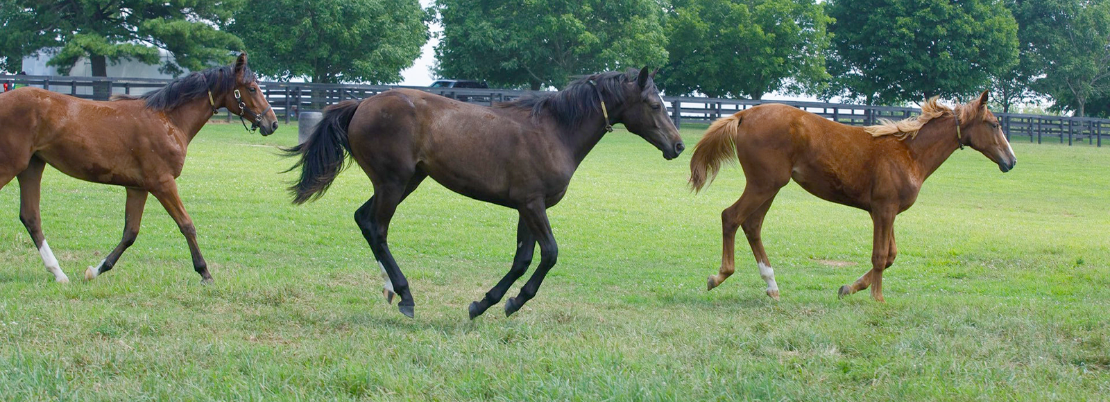
Prevention is key. Maintenance and early detection through an ongoing partnership with a licensed veterinarian will lead to optimal outcomes. Small problems that go undiagnosed or untreated may result in larger issues that could limit a horse’s performance career.
When a performance or training issue arises, start looking for a physical reason before a behavioral one. You’ll want to work with an experienced veterinarian who is familiar with your discipline and its common physical conditions. Many veterinarians who specialize in performance horse practice are familiar with multiple disciplines and skilled in treating horses that perform at the highest levels of competition.
Approximate Closure Times for Physes
- Birth: The physis at the top of the coffin bone within the hoof wall closes
- Birth – 6 months: The physes at the top and bottom of the short pastern bone close
- 6 months – 1 year: The physes at the top and bottom of the long pastern bone close
- 8 months – 1.5 years: The physes at the top and bottom of the cannon bone close
- 1.5 – 2.5 years: The small bones within the knee continue to develop and enlarge (some of the small bones in the hock continue to develop up until 4 years of age)
- 2 – 2.5 years: The physes at the bottom of the radius and ulna (forearm) close
- 2 – 3 years: Physes within the stifle close between 2 and 3 years of age
- 3 – 3.5 years:
- The physes at the top and bottom of the humerus close
- The physes at the top and bottom of the tibia close
- The physes at the bottom of the femur close
- Some of the physes in the pelvis do not close until 3 to 4 years of age
- Physes within the vertebrae of the spinal column may not be fully closed until 5 years of age. Time of closure may be longer in males and in the larger breeds
OCD lesions
OCD Lesions
OCD lesions, or osteochondritis dissecans, can be observed in yearlings. No one knows for sure why some horses develop OCD lesions, but genetics, environment, nutrition and amount of exercise have all been found to contribute to the condition. OCD lesions also are found more commonly in certain breeds such as Thoroughbreds, Standardbreds and Warmbloods, although any breed can develop the lesions.
OCD stands for “osteochondritis desicans” and has somewhat become slang for a broad array of disorders that can occur in the young growing horse skeleton. OCD is actually a very specific type of developmental problem that occurs at joint surfaces. What were often thought to be separate diagnostic issues in young horses are now thought to be closely related. Concerns like contracted tendons, epiphysitis, swollen joints and even wobbler syndrome may actually be closely related to problems that occur as the bones of young horses grow. A broad term that we use today is developmental orthopedic disease, or DOD.
Clinical Signs Include:
- Lameness
- Joint effusion (increased fluid in the joint)
- Some horses will show no sign of orthopedic disease until strenuous exercise is started
Radiographs are an excellent way of screening for these conditions in a young horse, especially those without clinical signs. The most common sites of OCD lesions are the stifle and hock, and, to a lesser extent, the fetlocks. Discuss with your veterinarian screening for OCD as part of your pre-purchase exam.
Splints
Splints occur most commonly in the growing horse in training. Splint bones (also known as the second and fourth metacarpal bones) are found on either side of the cannon bone (third metacarpal bone). Theses bones have a fibrous connective tissue structure called the interosseous ligament, which calcifies into bone and fuses with the adjacent cannon bone as the horse matures. Complete fusion usually occurs by the time the horse is around 3 to 4 years of age.
The condition of “splints” refers to an inflammation between the interosseous ligament and the splint bone itself or the soft tissue covering of the bone called the periosteum. This condition is usually the result of injury from external trauma or tearing of the interosseous ligament due to overtraining. Most splints occur on the inside, or medial side, of the front limbs a couple of inches from the knee or carpus. Lateral or outside splints are less common but can occur on either the front or hind limbs.
Risk Factors
The most common risk factor for splints is training/exercise. Heat, swelling, lameness and tenderness are the primary clinical signs. The lameness is usually mild and more evident at the trot or gallop.
Pain on palpation is almost always present, and radiographs can confirm the diagnosis as well as give an indication of the degree of the cosmetic prognosis. Radiographs also can rule out a splint bone fracture that could possibly require surgical intervention and a longer rehabilitation period. Ultrasound can be useful to determine the extent of any soft tissue damage.
Treatment
The treatment for splints is rest followed by a structured rehabilitation program that includes hand walking and a slow introduction back into work. Cold water therapy during the early stages of the condition and anti-inflammatory drugs such as Banamine® (flunixin meglumine paste) or phenylbutazone are helpful at decreasing the inflammation and swelling. Bandaging and other topical anti-inflammatory therapies also are beneficial.
Important Safety Information
BANAMINE Paste: For Oral Use in Horses Only. Not for use in horses intended for human consumption. Do not use in horses showing hypersensitivity to flunixin meglumine. The effect of BANAMINE Paste on pregnancy has not been determined. Concomitant use of Banamine with other anti-inflammatory drugs such as NSAIDs and corticosteroids should be avoided or closely monitored. For complete information on Banamine® Paste, see accompanying product package insert.
Other Concerns
Eye
Many vision problems can manifest as a training issue. That’s why we recommend a proper eye exam prior to breaking and training and as part of the pre-purchase examination.
Young horses need an annual physical including eye, dental and lameness exams, as well as body condition scoring and evaluation of skeletal and foot conformation.
Heart
Have your veterinarian listen to their heart for signs of underlying heart disease. This can be done at your horse’s annual physical exam.
If abnormalities are detected, your veterinarian will use more sensitive diagnostics such as ultrasound or echocardiogram. Loud murmurs and irregular heartbeats can potentially indicate underlying disease.
Reproductive
This is the age where you determine whether to leave a young horse intact or not. Have your veterinarian check to make sure both testicles are descended.
Fillies will come into heat for the first time at this age, so make sure they are housed separately from intact colts.
Skin Lesions
Warts commonly occur in growing horses.
Warts are caused by the papillomavirus and usually appear on the nose, muzzle or face of the horse. They can appear as single lesions or a cluster of lesions with a cauliflower appearance.
While they pose a cosmetic concern, warts rarely pose a significant health threat to the young horse that is properly maintained through good nutrition and management. The infections are self-limiting (resolve on their own), and after the episode has cleared, horses develop immunity to this virus so reoccurrences do not usually happen.
Treatment is not typically necessary unless the lesions become open and infected due to injury from tack, stall bars or doors. These secondary infections can be managed with topical therapy and daily cleaning. Some people feel that surgically removing the warts can speed up the healing and the immune process. This is controversial, however, since the infection resolves on its own. Typically, warts should clear up in six to nine months.