
Equine Health Library
Young Horse
Health Conditions
Neurologic | Gastrointestinal | Respiratory
Neurologic
Neurologic
Noninfectious conditions, such as cervical vertebral stenotic myelopathy, may be mistaken for infectious neurologic diseases including Equine Protozoal Myeloencephalitis (EPM), West Nile virus, Equine Herpesvirus (EHV) or rabies. Therapy and prognosis can vary greatly depending on the cause of neurologic disease.
Eastern Equine Encephalomyelitis (EEE)
Mosquitoes aren’t just annoying. They can carry viruses that cause neurological diseases like Eastern Equine Encephalomyelitis (EEE).
EEE is a virus that causes severe and often fatal neurologic disease in horses, humans and other mammals. The disease is found as far north as eastern Canada and as far south as Central and South America. In the United States, EEE has been detected in all states east of the Mississippi River, as well as a number of western states. Every year, several hundred horses are diagnosed with EEE. Young horses are most susceptible to infection.
Clinical Signs of EEE
- High fever
- Severe depression
- Incoordination
- Paralysis
- Seizures
- Coma
EEE is fatal in most horses. Prevention focuses on mosquito control and appropriate vaccination once or twice per year depending on the climate and other disease risk factors. EEE is an essential vaccine that all horses should receive. In areas with long mosquito seasons, horses are typically vaccinated twice a year against EEE. Vaccination against EEE is highly effective at preventing clinical disease.
Cervical Vertebral Stenotic Myelopathy
Cervical Vertebral Stenotic Myelopathy (CVSM), is a neurologic disease that often becomes apparent when young horses first enter training. It’s a result of cervical vertebrae compressing or impinging on the spinal cord as it descends along the neck. Young, large, rapidly growing horses (usually males) are most at risk for one form of this condition caused by narrowing of the cervical spinal canal. Movements that include twisting and turning of the horse’s long neck can exacerbate spinal cord compression due to instability between vertebrae.
Signs of CVSM Include:
- Incoordination (ataxia)
- Lack of awareness where the limbs are in space (proprioceptive deficits)
- Progressive clumsiness or weakness characterized by knuckling, stumbling or dragging the limbs, incoordination and spasticity
- Hind limbs are often affected before the forelimbs
- Among young growing horses, Thoroughbreds, Quarter Horses and Warmbloods are the more commonly affected breeds
- Colts and stallions are more likely to develop the syndrome
Causes of CVSM
In addition to genetics, rapid growth can be a contributing factor. Trauma, such as a fall or rough play at pasture, also can precipitate the sudden onset of neurologic signs.
Diagnosis and Treatment of CVSM
Diagnosis requires a thorough physical and neurological examination to rule out other causes of neurologic disease and lameness. Standing radiographs of the neck can help identify unusually narrow segments of the spinal canal. A myelogram (injection of contrast dye into the spinal fluid surrounding the spinal cord) may be recommended to pinpoint specific areas of spinal cord impingement.
Severe cases of CVSM can render the horse a danger to himself and those around him. Milder cases can be helped with surgical procedures designed to stabilize unstable vertebrae. Anti-inflammatory medications and rest can help relieve the symptoms.
Gastrointestinal
Gastrointestinal
Young horses with immature immune systems are more susceptible to parasitism than older horses. Ascarids, tapeworms, and cyathostomes (small strongyles) are three parasites that can cause colic.
Colic in Young Horses

Colicky foal.
Behavior to Watch For
Young foals are often very demonstrative when in pain regardless of the cause. The amount of kicking and rolling often cannot be correlated with the severity of the abdominal problem.
A foal with a meconium impaction that is relieved by an enema and a foal with a more serious intestinal problem requiring surgery may be difficult to distinguish from one another at first glance. Therefore, every episode of colic in a foal should be carefully evaluated by your veterinarian.
Parasites that Cause Colic
- Ascarids — Heavy burdens of maturing ascarid larvae and adults can cause small intestinal impaction or rupture – with secondary peritonitis (inflammation of the thin tissue that lines the inner wall of the abdomen and covers most of the abdominal organs). Recent deworming may precede colic and is believed to be due to suddenly killing large numbers of ascarids. If heavy ascarid burdens are suspected, talk with your veterinarian before administering any dewormer. Youngsters harboring large numbers of ascarids may benefit from being pre-treated with Banamine® (flunixin meglumine paste). Because of its method of killing ascarids, fenbendazole may be one of the safer dewormers to use under these conditions.
Click here to learn more about Ascarids. - Tapeworms — Adult tapeworms cluster and attach to the mucosa lining of the distal small intestine and ileocecal valve (connection between the end of the small intestine, called the ileum, and the cecum). Tapeworms have been associated with ileocecal intussusceptions, ileal impactions and some cases of spasmodic colic. Tapeworm eggs can be difficult to detect in fecal exams. A double dose of pyrantel or a dewormer containing praziquantel are the two treatment options for tapeworm infections.
- Small Strongyles — Encysted small strongyles (cyathostomes) can cause colic, weight loss and diarrhea when large numbers of encysted larvae begin to emerge spontaneously from their site of hibernation within the wall of the large intestine and re-enter the lumen of the bowel. Larvicidal fenbendazole (PANACUR® (fenbendazole) POWERPAC) and moxidectin are the only dewormers labeled to treat encysted stages of small strongyles.
Click here to learn more about small strongyles.
Important Safety Information
BANAMINE Paste: For Oral Use in Horses Only. Not for use in horses intended for human consumption. Do not use in horses showing hypersensitivity to flunixin meglumine. The effect of BANAMINE Paste on pregnancy has not been determined. Concomitant use of Banamine with other anti-inflammatory drugs such as NSAIDs and corticosteroids should be avoided or closely monitored. For complete information on Banamine® Paste, see accompanying product package insert.
PANACUR® (fenbendazole) POWERPAC: NOT FOR USE IN HUMANS. KEEP OUT OF REACH OF CHILDREN. Do not use in horses intended for human consumption.
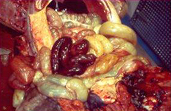
Volvulus (torsion) of the small intestines in a young foal with colic. The discolored loops of bowel are the result of complete obstruction of blood flow to the affected loops of bowel.
Ulcers Can Cause Colic
Gastric ulcers can be a source of colic symptoms, poor appetite and loss of condition. A risk factor for ulcers is stress – including stress of entering the training barn and beginning work, stress associated with shipping and stress associated with diet changes.
Respiratory
Respiratory
Infectious respiratory disease due to Equine Influenza, Equine Herpesvirus or Strangles is more common among young horses less than 5 years of age.
Environmental Factors
Equine asthma can occur in young horses recovering from viral respiratory diseases and can be exacerbated by sub-optimal barn ventilation, air pollutants, exposure to dusty bedding and/or moldy hay, and premature return to exercise after infection.
Talk to your veterinarian if your horse has trouble breathing or exhibits an increase in coughing or nasal discharge.
Equine Influenza Virus (EIV)
Equine influenza (EIV) is one of the most common infectious upper respiratory diseases in young horses. Influenza can spread quickly because the incubation period is only 24 hours to three days, and the virus can be transmitted through the air. In fact, coughing can spread nasal droplets more than 200 yards. Horses that are sick for the first time can shed the virus in nasal secretions for as long as seven to 10 days. Indirect transmission of the virus can also occur via hands, clothing and common use articles such as bits, brushes and buckets. Vaccination is the best prevention.
Clinical Signs Include
- High fever (>103 to 105 degrees Fahrenheit)
- Depression
- Dry hacking cough
- Clear nasal discharge (that becomes thick and colored if a secondary bacterial infection develops)
- Loss of appetite with secondary weight loss
- Muscle soreness
- Less common signs include distal limb edema and a rare form of heart disease (cardiomyopathy). More severe signs are observed in donkeys and mules with fatalities reported
These signs typically resolve in seven to 14 days, although the cough may persist for 21 days.
Diagnosis and Treatment
The fastest way to diagnose EIV is testing using nasal swabs. Recovering horses require a minimum of three weeks of rest or at least one week of rest for every day of fever. Premature return to exercise may be associated with complications including secondary bacterial infections, reactive airway disease and exercise intolerance.
Prevention
Vaccination is the best way to prevent influenza. EIV is considered an at-risk vaccination, and horses at risk for EIV include all young horses less than 4 to 5 years of age, horses in contact with large groups of horses (e.g., at shows, on trail rides, at riding clinics, etc.) and stay-at-home horses on farms where other resident horses are going to and coming from events and shows. Mature, healthy horses, in general, are less susceptible. Most horses should be vaccinated against EIV unless they live in a closed herd or isolated facility. Frequency of vaccination is determined by risk and type of vaccine.
Biosecurity is another critical step in the prevention of influenza.
Action Items
- Isolate new arrivals for two to three weeks and observe for fever, cough or nasal discharge
- Monitor the daily temperature of all incoming horses for at least the first three to five days following their arrival and at the first sign of any respiratory disease or loss of appetite.
Equine Herpesvirus (EHV 1 & 4)
Equine herpesvirus type 1 (EHV-1) and equine herpesvirus type 4 (EHV-4) can each infect the respiratory tract, causing disease that varies in severity from subclinical (not apparent) to severe. EHV-4 is typically associated with upper respiratory disease in younger horses; while EHV-1 can cause respiratory disease, late-term abortions, early foal deaths and/or neurologic disease.
Most horses have been infected with EHV-1 by the time they are yearlings. After initial infection, equine herpesvirus remains dormant (latent) in the horse. Stressful events, such as hauling, handling and training can reactivate the virus, and viral shedding can occur “silently” – without symptoms.
How it’s Spread
The incubation period for EHV-1 is four to six days but can be as short as 24 hours or as long as 10 days. Clinical signs are typically observed within one to three days. Shedding of the virus typically lasts seven to 10 days but can last as long as 28 days. Longer shedding periods and higher viral loads have been reported in horses experiencing the neurologic form of the disease, Equine Herpesvirus Myeloencephalopathy (EHM).
Clinical signs of respiratory disease vary depending on the virus strain and immune status of the horse:
- Two fever spikes – one to two days after infection and six to seven days after infection
- Depression
- Loss of appetite
- Cough (less common)
- Clear nasal discharge (may become thick and colored if a secondary bacterial infection occurs)
- Conjunctivitis (reddening around the eyes)
- Mild to moderate lymph node enlargement that can persist for weeks
- Following recovery some horses develop “poor performance syndrome” and reactive airway disease
To diagnose EHV-1 infection, your veterinarian will submit a nasal swab and blood sample for PCR testing. Prevention of respiratory disease includes vaccination and appropriate biosecurity protocols. Currently, there is no vaccine labeled for the prevention of EHM.
Strangles
Strangles is a highly contagious disease caused by the abscess-forming, gram positive bacteria Streptococcus equi. Approximately 75 percent of recovered horses develop immunity for five years or longer. Asymptomatic horses (silent shedders) can harbor the bacteria in their guttural pouches for several years and represent a source of persistent infection. The bacteria itself is hardy and survives in a moist environment and organic debris such as manure for up to six to seven weeks. Direct sunlight shortens this time period considerably.
Clinical Signs
- High fever (≥ 103 degrees Fahrenheit). Fever is usually the first sign of disease and precedes other symptoms by 24 to 48 hours
- Thick pus-like nasal discharge. Nasal shedding of bacteria begins one to two days after the onset of fever and can persist for two to three weeks
- Enlarged, swollen and tender lymph nodes around the head, under the jaw and around the throat latch that frequently abscess, rupture and drain. Abscesses can also develop on other places of the body, both externally and internally. (Internal abscesses are called “bastard strangles” and are much more difficult to treat)
- Difficulty swallowing and breathing due to enlarged lymph nodes placing pressure around the back of the throat, the esophagus and the trachea
- Loss of appetite
- Depression
- Occasional soft moist cough
Diagnosis and Treatment
Your veterinarian can confirm the diagnosis of strangles using nasal swabs, nasal and guttural pouch washes and/or pus aspirated from abscesses and submitted for culture and testing. A persistent infection in a horse’s guttural pouch is best visualized using an endoscope. This is a known method for identifying horses that are silent carriers of the disease.
Once external abscesses “mature,” they can be opened, drained and flushed. Some abscesses will rupture spontaneously. Anti-inflammatory drugs such as Banamine® (flunixin meglumine paste) may be administered as needed to keep horses comfortable enough to continue eating and drinking. Discuss all medications with your veterinarian. External abscesses often require many weeks to completely resolve, and while they do, the draining pus is highly contagious. Anyone handling affected horses should wear disposable gloves and should not handle uninfected horses without first changing their clothes and washing their hands.
Internal abscesses (or “bastard strangles”) can develop in internal lymph nodes as well as in other organs. Some common internal locations include: The lungs, liver, spleen, kidney, brain and the lymph nodes lining the intestinal tract. Internal abscesses require prolonged, systemic antibiotic therapy lasting several months, and even then abscesses may not fully resolve.
Purpura hemorrhagica is an uncommon complication that can occur in horses recovering from natural infection with Streptococci equi and occasionally in horses following vaccination against strangles. Purpura is an immune-mediated inflammation of the lining of blood vessels. Affected horses develop severe, pitting edema along all dependent areas of the body: Under the jaw, along the ventral abdomen and in all the limbs. Often the skin covering the edematous areas begins to ooze serum. Skin may even slough over affected regions. Purpura hemorrhagica can be life-threatening and requires aggressive treatment with antibiotics, steroids and anti-inflammatory drugs in addition to good nursing care.
Strangles Prevention
Prevention and containment of the disease should focus on hygiene, disinfection and identification of silent shedders. Horses can acquire strangles through both horse-to-horse contact and indirect transmission via contaminated shared equipment (stalls, water buckets and troughs, feeding tubs, twitches, clothing, handlers’ hands, etc.). A good biosecurity program is vital to reducing the risk of a strangles outbreak. Quarantine new arrivals for 21 to 28 days. Your veterinarian will likely recommend screening suspects or recovering horses with weekly nasal or guttural pouch washes and keep affected horses isolated until three negative cultures and PCR tests are obtained.
Your veterinarian can help design a practical biosecurity program for your operation to decrease the likelihood that your horses will contract this disease in the future. Remember, outwardly normal horses that have recovered from an episode of strangles can be the silent shedders that begin the next outbreak. Work with your veterinarian to identify those shedders and treat them. Quarantine all new arrivals for at least two to three weeks to help prevent the introduction of strangles and other contagious diseases.
Intramuscular killed vaccines, and an intranasal modified live vaccine, are available in the U.S. Vaccination during an outbreak is generally not recommended unless it is certain that the horses to be vaccinated are not incubating the disease and do not have exceptionally high antibody titers against S. equi. Your veterinarian can screen your horse for pre-existing S. equi antibodies if this is a concern.
Important Safety Information
BANAMINE Paste: For Oral Use in Horses Only. Not for use in horses intended for human consumption. Do not use in horses showing hypersensitivity to flunixin meglumine. The effect of BANAMINE Paste on pregnancy has not been determined. Concomitant use of Banamine with other anti-inflammatory drugs such as NSAIDs and corticosteroids should be avoided or closely monitored. For complete information on Banamine® Paste, see accompanying product package insert.